




Prevention and Treatment of Refractive Errors
During every summer holiday
many parents take their children to ophthalmology clinics
Among these students, the majority have vision problems
with refractive errors being a relatively common eye condition
With the widespread use of electronic devices
students are spending increasing amounts of time on screens
leading to greater visual strain
This has caused the incidence of refractive errors to rise
bringing considerable inconvenience to students' studies and daily lives
Therefore
the prevention and treatment of refractive errors are particularly important
Next
experts from the Department of Ophthalmology at the Army Special Medical Center (Daping Hospital) of the Army Medical University
will explain the relevant knowledge
I. What is a Refractive Error?
When the eye's accommodation is at rest, parallel light rays from the outside pass through the eye's refractive system but fail to focus on the fovea centralis of the retina, thus unable to produce a clear image. This condition is called a refractive error. Refractive errors include myopia, hyperopia, and astigmatism.
Myopia: Myopia occurs when the axial length of the eye is too long or the corneal refractive power is too strong, causing parallel light rays entering the eye to focus in front of the retina rather than on it, resulting in a blurred image.
Based on the degree of myopia, it can be classified into mild, moderate, high, and very high myopia. Mild myopia is up to -3.00 D, moderate myopia is between -3.00 D and -6.00 D, high myopia is between -6.00 D and -10.00 D, and very high myopia is over -10.00 D.
Furthermore, myopia can be divided into simple myopia and pathological myopia. Patients with pathological myopia have unstable prescriptions and are prone to complications.
Hyperopia: Hyperopia occurs when the anteroposterior diameter of the eyeball is too short or the refractive power of the eye's optical system is insufficient, causing parallel light beams entering the eye from the front to focus behind the retina, resulting in a blurred image. This condition is known as hyperopia.
Hyperopia can be classified by degree as follows:
- Low hyperopia: < +3.00 D. In youth, since accommodation can compensate for distance vision, most people's vision is unaffected before the age of 40.
- Moderate hyperopia: +3.00 D to +5.00 D. Vision is affected at this stage, accompanied by discomfort or asthenopia symptoms. Excessive use of accommodation may also lead to esotropia.
- High hyperopia: > +5.00 D. Vision is significantly affected and objects appear blurred, but visual fatigue or discomfort is not prominent. This is because the hyperopia is so high that the patient cannot compensate through accommodation.
Astigmatism: Astigmatism is closely related to the curvature of the cornea. When parallel light rays enter the eye, due to differences in refractive power across different meridians of the eyeball, they cannot focus on a single point (i.e., a focal point), thus failing to form a clear image. This condition is called astigmatism. In an eye with astigmatism, neither accommodation nor changing the distance between the target and the eye can produce a clear image. Only by wearing appropriate cylindrical lenses can a clear image be formed on the retina.
II. How to Prevent Refractive Errors?
Students can take the following measures in their daily life and studies to protect their vision:
Reasonably control the use of electronic devices; each session should not exceed half an hour, followed by looking into the distance to relax the eyes.
When reading and writing, keep a distance of about 33 centimetres between the eyes and the book, one fist between the chest and the desk, and one inch between the fingers holding the pen and the tip of the pen.
Ensure at least one hour of outdoor activity every day to allow the eyes to be fully exposed to natural light.
Undergo regular vision checks to identify and address any issues promptly.
III. How to Correct Refractive Errors?
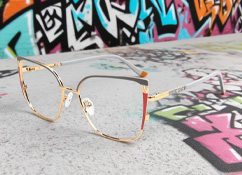
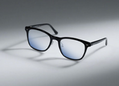
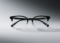
Refractive errors can be corrected by wearing spectacles or through refractive surgery. The current mainstream refractive surgery methods include:
- Excimer laser surgery
By removing the corneal epithelium through physicochemical means, the cornea is ablated with an excimer laser. This maximises the preservation of corneal tissue and maintains corneal biomechanical properties, thereby improving surgical safety.
- Femtosecond laser-assisted LASIK (FS-LASIK)
This uses a femtosecond laser instead of a traditional mechanical microkeratome to create the corneal flap. After the flap is created, the corneal stroma is ablated using an excimer laser. This procedure avoids the trauma caused by a mechanical blade, making the surgery safer and more precise. It has a lesser impact on corneal biomechanics and allows for customised parameter design, resulting in better postoperative outcomes.
- Small Incision Lenticule Extraction (SMILE)
The surgery directly uses a femtosecond laser to create a lenticule within the corneal stroma, which is then extracted through a small incision. This procedure has a minimal impact on corneal biomechanics and avoids the risk of corneal flap displacement.
- Implantable Collamer Lens (ICL) Implantation
ICL involves implanting a custom-prescribed soft lens inside the eye to correct vision. Its main advantages are that the procedure is reversible, requires no corneal tissue removal, and offers a wide range of correction.
ICL intraocular lens implantation surgery
IV. Frequently Asked Questions About Refractive Surgery
- Q: What should I do if I have myopia? Do I have to wear glasses for the rest of my life?
A: You can wait until you are 18 years old and your prescription has stabilised before undergoing myopia surgery.
- Q: Who is suitable for myopia surgery?
A: Suitable candidates must have a clear desire to be free from glasses, be at least 18 years old, and have had a stable prescription for the past two years.
Corneal refractive surgery: requires a corneal thickness of no less than 470 micrometres; myopia between -1.00 D and -10.00 D, hyperopia up to 6.00 D, and astigmatism up to 6.00 D.
Implantable Collamer Lens (ICL) Implantation: suitable for myopia between -1.00 D and -18.00 D, and astigmatism up to 6.00 D.
Contact lens wearers should stop wearing soft lenses for 2 weeks and rigid lenses for at least 4 weeks prior. Additionally, eye examinations must show no abnormalities, no active eye diseases, and no autoimmune diseases.
- Q: Will I no longer be myopic after the surgery?
A: Myopia surgery is merely a "symptomatic" procedure to eliminate the need for glasses; it does not cure myopia itself. Therefore, you must continue to protect your eyes postoperatively. Remember the "20-20-20" rule: after 20 minutes of near work (such as reading, writing, or looking at a mobile phone), look at an object at least 20 feet (about 6 metres) away for at least 20 seconds.
- Q: Does myopia surgery hurt?
A: Although myopia surgery is performed while the patient is fully awake, anaesthetic eye drops are administered beforehand, so there is no significant pain during the procedure.
- Q: Is it difficult to cooperate during refractive surgery?
A: Refractive surgery requires the patient to cooperate with the surgeon during the procedure; throughout the process, you only need to fixate on the green guiding light. SMILE requires the patient to keep their eyes open and still for 30 seconds, while customised femtosecond LASIK requires 15 seconds. There is no need to worry about blinking, as after the anaesthetic drops are administered, everyone can keep their eyes open without blinking for over 2 minutes.
- Q: Can I exercise after refractive surgery?
A: Within the first three days post-surgery, you may engage in light exercises such as jogging, walking, and yoga. After three days, you can resume various fitness and physical training activities; however, swimming should be postponed until three months after the surgery.